|
R&D Systems
anti goat igg hrp 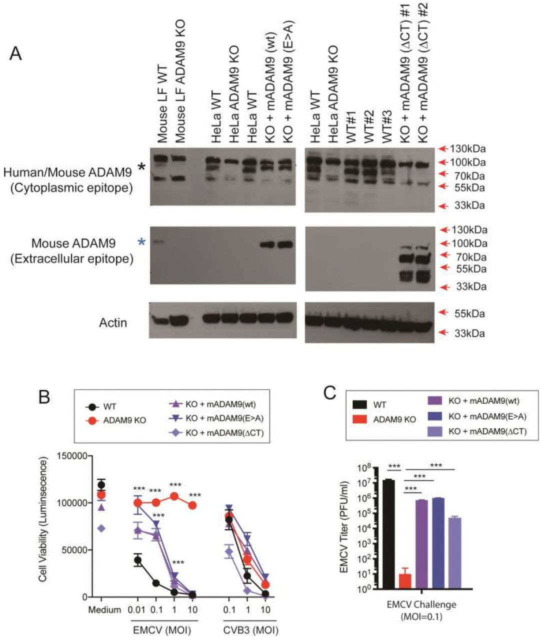 Anti Goat Igg Hrp, supplied by R&D Systems, used in various techniques. Bioz Stars score: 99/100, based on 1 PubMed citations. ZERO BIAS - scores, article reviews, protocol conditions and more https://www.bioz.com/product/hrp+conj/Goat+IgG+HRP-conjugated+Antibody/bio_rxiv__491068-209-4-9 Average 99 stars, based on 1 article reviews
anti goat igg hrp - by Bioz Stars,
2026-08
99/100 stars
|
Buy from Supplier |
|
R&D Systems
rabbit anti goat igg conjugated horseradish peroxidase Rabbit Anti Goat Igg Conjugated Horseradish Peroxidase, supplied by R&D Systems, used in various techniques. Bioz Stars score: 96/100, based on 1 PubMed citations. ZERO BIAS - scores, article reviews, protocol conditions and more https://www.bioz.com/product/hrp+conj/Goat+IgG+HRP-conjugated+Antibody/pm27815474-95-30-37 Average 96 stars, based on 1 article reviews
rabbit anti goat igg conjugated horseradish peroxidase - by Bioz Stars,
2026-08
96/100 stars
|
Buy from Supplier |
|
Boster Bio
horseradish peroxidase conjugated goat Horseradish Peroxidase Conjugated Goat, supplied by Boster Bio, used in various techniques. Bioz Stars score: 96/100, based on 1 PubMed citations. ZERO BIAS - scores, article reviews, protocol conditions and more https://www.bioz.com/product/hrp+conj/HRP+Conjugated+AffiniPure+Goat+Anti-Rabbit+IgG/pm41804010-72-12-19 Average 96 stars, based on 1 article reviews
horseradish peroxidase conjugated goat - by Bioz Stars,
2026-08
96/100 stars
|
Buy from Supplier |
|
R&D Systems
mouse monoclonal anti phospho tyrosine hrp conjugated ab Mouse Monoclonal Anti Phospho Tyrosine Hrp Conjugated Ab, supplied by R&D Systems, used in various techniques. Bioz Stars score: 93/100, based on 1 PubMed citations. ZERO BIAS - scores, article reviews, protocol conditions and more https://www.bioz.com/product/hrp+conj/Phospho-Tyrosine+HRP-conjugated+Antibody/pm33452125-55-35-43 Average 93 stars, based on 1 article reviews
mouse monoclonal anti phospho tyrosine hrp conjugated ab - by Bioz Stars,
2026-08
93/100 stars
|
Buy from Supplier |
|
R&D Systems
donkey anti goat igg hrp conjugated antibody Donkey Anti Goat Igg Hrp Conjugated Antibody, supplied by R&D Systems, used in various techniques. Bioz Stars score: 94/100, based on 1 PubMed citations. ZERO BIAS - scores, article reviews, protocol conditions and more https://www.bioz.com/product/hrp+conj/Goat+IgG+HRP-conjugated+Antibody/pm39548070-551-28-33 Average 94 stars, based on 1 article reviews
donkey anti goat igg hrp conjugated antibody - by Bioz Stars,
2026-08
94/100 stars
|
Buy from Supplier |
|
R&D Systems
monoclonal mouse igg hrp conjugated anti6xhis antibody Monoclonal Mouse Igg Hrp Conjugated Anti6xhis Antibody, supplied by R&D Systems, used in various techniques. Bioz Stars score: 99/100, based on 1 PubMed citations. ZERO BIAS - scores, article reviews, protocol conditions and more https://www.bioz.com/product/hrp+conj/Mouse+IgG+HRP-conjugated+Antibody/pm24036079-71-1-9 Average 99 stars, based on 1 article reviews
monoclonal mouse igg hrp conjugated anti6xhis antibody - by Bioz Stars,
2026-08
99/100 stars
|
Buy from Supplier |
|
Bethyl
light chain cross adsorbed antibody hrp conjugated Light Chain Cross Adsorbed Antibody Hrp Conjugated, supplied by Bethyl, used in various techniques. Bioz Stars score: 95/100, based on 1 PubMed citations. ZERO BIAS - scores, article reviews, protocol conditions and more https://www.bioz.com/product/hrp+conj/Rabbit+Light+Chain+Antibody+HRP+Conjugated/pmc09644419-190-8-13 Average 95 stars, based on 1 article reviews
light chain cross adsorbed antibody hrp conjugated - by Bioz Stars,
2026-08
95/100 stars
|
Buy from Supplier |
|
Proteintech
ferritin heavy chain polyclonal antibody fth1  Ferritin Heavy Chain Polyclonal Antibody Fth1, supplied by Proteintech, used in various techniques. Bioz Stars score: 96/100, based on 1 PubMed citations. ZERO BIAS - scores, article reviews, protocol conditions and more https://www.bioz.com/product/hrp+conj/HRP-conjugated+Heavy+chain+of+Rabbit+IgG+Antibody/pm39744125-73-28-34 Average 96 stars, based on 1 article reviews
ferritin heavy chain polyclonal antibody fth1 - by Bioz Stars,
2026-08
96/100 stars
|
Buy from Supplier |
|
Proteintech
anti gfp Anti Gfp, supplied by Proteintech, used in various techniques. Bioz Stars score: 94/100, based on 1 PubMed citations. ZERO BIAS - scores, article reviews, protocol conditions and more https://www.bioz.com/product/hrp+conj/HRP-conjugated+GFP+tag+Antibody/pm40221426-470-17-18 Average 94 stars, based on 1 article reviews
anti gfp - by Bioz Stars,
2026-08
94/100 stars
|
Buy from Supplier |
|
Proteintech
β actin  β Actin, supplied by Proteintech, used in various techniques. Bioz Stars score: 96/100, based on 1 PubMed citations. ZERO BIAS - scores, article reviews, protocol conditions and more https://www.bioz.com/product/hrp+conj/HRP-conjugated+beta+Actin+Monoclonal+antibody/pmc12934520-59-74-91 Average 96 stars, based on 1 article reviews
β actin - by Bioz Stars,
2026-08
96/100 stars
|
Buy from Supplier |
|
Proteintech
anti gapdh Anti Gapdh, supplied by Proteintech, used in various techniques. Bioz Stars score: 96/100, based on 1 PubMed citations. ZERO BIAS - scores, article reviews, protocol conditions and more https://www.bioz.com/product/hrp+conj/HRP-conjugated+GAPDH+Monoclonal+antibody/pm41894180-39-39-43 Average 96 stars, based on 1 article reviews
anti gapdh - by Bioz Stars,
2026-08
96/100 stars
|
Buy from Supplier |
|
Proteintech
hrp conjugated affinipure goat antimouse igg Hrp Conjugated Affinipure Goat Antimouse Igg, supplied by Proteintech, used in various techniques. Bioz Stars score: 94/100, based on 1 PubMed citations. ZERO BIAS - scores, article reviews, protocol conditions and more https://www.bioz.com/product/hrp+conj/HRP-conjugated+Affinipure+Goat+Anti-Horse+IgG/pm36851019-90-100-109 Average 94 stars, based on 1 article reviews
hrp conjugated affinipure goat antimouse igg - by Bioz Stars,
2026-08
94/100 stars
|
Buy from Supplier |
Image Search Results
Journal: bioRxiv
Article Title: Identification of A Disintegrin and Metalloproteinase 9 domain (ADAM9) required in the early stages of encephalomyocarditis virus infection
doi: 10.1101/491068
Figure Lengend Snippet: WT and ADAM9 KO HeLa cells were transduced with retroviral vectors with wild-type (WT) murine ADAM9 (mADAM9), catalytically inactive mutant ADAM9 (E>A), cytoplasmic tail deleted (ΔCT) ADAM9 constructs, or GFP control vectors. Transduced cells were selected and cloned by limiting dilution, and expression of mADAM9 was confirmed by western blot analysis. (A) WT, KO and rescue cell lysates were prepared in RIPA buffer and run on 12% SDS-PAGE gels and transferred to PVDF membranes. Membranes were blocked with 3% BSA and incubated with ADAM9 antibodies. Upper panel, rabbit anti-human ADAM9 (Cell signaling #2099) that detects an epitope in the intracellular domain of human ADAM9 and cross-reacts with mouse ADAM9. Middle panel, goat anti-mouse ADAM9 (R&D systems AF949) that detects the extracellular domain of murine ADAM9, but not human ADAM9. Lower panel, anti-Actin (Santa Cruz sc-1616) which detects both human and murine ϟ-Actin. Bands were visualized using HRP and ECL reagent. Upper panel: WT but not KO cells expressed human ADAM9. Middle panel: Rescue but not KO cells expressed murine ADAM9. Lower panel: Actin loading control. (B) WT clones, ADAM9 KO clones and rescued ADAM9-expressing clones were infected with EMCV or CVB3 at varying MOI and incubated at 37 °C for 24 h. Viability of EMCV-infected and CVB3-infected clones was measured by CellGlo ATP luminescence. (C) EMCV replication was quantified in infected culture supernatants by plaque assay using BHK-21 cells. Neither the functional sequence of the ADAM9 metalloproteinase domain nor the cytoplasmic tail are required for EMCV infection. ***, P <0.0001, KO vs. WT; KO vs. rescue.
Article Snippet: 3% BSA blocking buffer),
Techniques: Transduction, Mutagenesis, Construct, Clone Assay, Expressing, Western Blot, SDS Page, Incubation, Infection, Plaque Assay, Functional Assay, Sequencing
Journal: Frontiers in pharmacology
Article Title: Transcription factor MEF2D regulates aberrant expression of ACSL3 and enhances sorafenib resistance by inhibiting ferroptosis in HCC.
doi: 10.3389/fphar.2024.1464852
Figure Lengend Snippet: FIGURE 2 The aberrant expression of ACSL3 in HCC protects cells from ferroptosis. (A) Genes highly correlated with ACSL3 in HCC were analyzed using Pearson text (B, C) Heatmaps displayed the top 50 genes that are positively and negatively correlated with ACSL3 in HCC. (D) KEGG pathway analysis revealed the correlation between ACSL3 and fatty acid biosynthesis and ferroptosis signaling pathway (E, F) After transfecting ACSL3-oe and control plasmids into PLC/PRF/5 cells, the mRNA and protein expression levels of ACSL4, GPX4, and FTH1 were quantified using qPCR and Western blotting. Vinculin protein served as the internal control. ****p < 0.0001, ***p < 0.001, **p < 0.01, *p < 0.05.
Article Snippet: ACSL3 antibody (Santa cruz, 1:100, Cat# sc-166374), ACSL4 Rabbit pAb (ABclonal, 1:1,000, Cat#A6826), MEF2D antibody (Santa cruz, 1:500, Cat# sc-271153), GPX4Monoclonal antibody (proteintech, 1:1,000, Cat No.: 67763-1- Ig),
Techniques: Expressing, Control, Western Blot
Journal: Investigative Ophthalmology & Visual Science
Article Title: AMPK Deficiency Induces Corneal Epithelial Barrier Dysfunction by Modulating Energy Homeostasis
doi: 10.1167/iovs.67.2.47
Figure Lengend Snippet: Construction and verification of AMPKα1α2CE-KO mice. (A) Breeding strategy. Cre recombinase excised the loxP-flanked exon 3 of AMPKα1 and exon 2 of AMPKα2; (B) Representative agarose gels showing the genotyping of wild type (WT) mice and AMPKα 1 α 2 CE-KO KO mice; PCR was performed on tail genomic DNA using primers (shown in red ), generating products of 334 bp and 341 bp from the WT allele, 450 bp from the floxed allele; (C) Representative images of immunofluorescence staining for AMPK ( red ) and K12 ( green ) in cornea of AMPKα1α2 f/f (Flox) and KO mice. Scale bar : 50 µm. (D) The immunofluorescence intensity of AMPKα in corneal epithelium. (E, F) Western blot and quantitative analysis of protein expression of p-AMPKα, AMPKα, and p-ACC in corneal epithelium with β-actin as a loading control. n = 3 (B–D) , n = 5 (E and F) . Data shown as mean ± SD. *** P < 0.001.
Article Snippet: Primary antibodies used in this study were as follows: p-AMPKα (2535, 1:1000), AMPKα (5831, 1:1000), phosphorylated acetyl-CoA carboxylase (p-ACC, 11818, 1:1000), ACC (3676, 1:1000), and Dynamin-related protein 1 (DRP1; 5391, 1:1000) from Cell Signaling Technology (Danvers, MA, USA); ZO-1 (21773-1-AP, 1:1000), Occludin (66378-1-lg, 1:1000), E-cadherin (CDH, 20874-1-AP, 1:1000), Mitofusion 2 (MFN2, 12186-1-AP, 1:1000), Optic atrophy 1 (OPA1, 27733-1-AP, 1:1000), MFF (17090-1-AP, 1:1000), IL-1β (16806-1-AP, 1:1000), IL-10 (60269-1-lg, 1:1000), Mitochondria Complex IV (MTCO2, 55070-1-AP, 1:1000),
Techniques: Immunofluorescence, Staining, Western Blot, Expressing, Control

Journal: Investigative Ophthalmology & Visual Science
Article Title: AMPK Deficiency Induces Corneal Epithelial Barrier Dysfunction by Modulating Energy Homeostasis
doi: 10.1167/iovs.67.2.47
Figure Lengend Snippet: AMPK deficiency induces disruption of epithelial AJCs in vivo and in vitro. (A, B) Western blot and quantitative analysis of protein expression of ZO1, Occludin, and CDH1 in corneal epithelium with β-actin as a loading control. (C) Representative images of immunofluorescence staining for ZO1 ( green ) in the cornea of Flox and KO mice; Scale bar : 50 µm. (D) The immunofluorescence intensity of ZO1 in corneal epithelium. (E) The mRNA expression of AMPKα1 and AMPKα2 in HCECs transfected with siCtrl and siAMPK. (F) Western blot and quantitative analysis of protein expression of AMPKα in HCECs with β-actin as a loading control. (G) Representative images of immunofluorescence staining for ZO1 ( green ) in HCECs. Scale bar : 25 µm. (H) Western blot and quantitative analysis of protein expression of ZO1, Occludin, and CDH1 in HCECs with β-actin as a loading control. n = 4. Data was shown as mean± SD. * P < 0.05, *** P < 0.001.
Article Snippet: Primary antibodies used in this study were as follows: p-AMPKα (2535, 1:1000), AMPKα (5831, 1:1000), phosphorylated acetyl-CoA carboxylase (p-ACC, 11818, 1:1000), ACC (3676, 1:1000), and Dynamin-related protein 1 (DRP1; 5391, 1:1000) from Cell Signaling Technology (Danvers, MA, USA); ZO-1 (21773-1-AP, 1:1000), Occludin (66378-1-lg, 1:1000), E-cadherin (CDH, 20874-1-AP, 1:1000), Mitofusion 2 (MFN2, 12186-1-AP, 1:1000), Optic atrophy 1 (OPA1, 27733-1-AP, 1:1000), MFF (17090-1-AP, 1:1000), IL-1β (16806-1-AP, 1:1000), IL-10 (60269-1-lg, 1:1000), Mitochondria Complex IV (MTCO2, 55070-1-AP, 1:1000),
Techniques: Disruption, In Vivo, In Vitro, Western Blot, Expressing, Control, Immunofluorescence, Staining, Transfection

Journal: Investigative Ophthalmology & Visual Science
Article Title: AMPK Deficiency Induces Corneal Epithelial Barrier Dysfunction by Modulating Energy Homeostasis
doi: 10.1167/iovs.67.2.47
Figure Lengend Snippet: AMPK deficiency impairs mitochondrial homeostasis in corneal epithelial cells. (A) Representative images of Rhodamine ( green ) staining in HCECs. Scale bar : 100 µm. (B) The fluorescence intensity of Rhodamine staining in HCECs. (C) OCR in siAMPK transfected HCECs versus siCtrl transfected HCECs. (D) Quantification of basal respiration, ATP production and maximal mitochondrial respiratory of HCECs in (C) . (E, F) Representative flow cytometry analysis images and quantification of MitoTracker Green staining in HCECs transfected with siCtrl and siAMPK. (G–I) Western blot and quantitative analysis of protein expression of MFN2, OPA1, DRP1, MFF, and Complex IV in HCECs with β-actin as a loading control. (J) Measurement of ATP levels in corneal epithelium of Flox and KO mice. (K–M) Western blot and quantitative analysis of protein expression of MFN2, OPA1, DRP1, MFF, and Complex IV in corneal epithelium with β-actin as a loading control. n = 6 (A–D) , n = 4 (G–I) , n = 3 (E, F, J, K–M) . Data shown as mean ± SD. * P < 0.05, ** P < 0.01, *** P < 0.001.
Article Snippet: Primary antibodies used in this study were as follows: p-AMPKα (2535, 1:1000), AMPKα (5831, 1:1000), phosphorylated acetyl-CoA carboxylase (p-ACC, 11818, 1:1000), ACC (3676, 1:1000), and Dynamin-related protein 1 (DRP1; 5391, 1:1000) from Cell Signaling Technology (Danvers, MA, USA); ZO-1 (21773-1-AP, 1:1000), Occludin (66378-1-lg, 1:1000), E-cadherin (CDH, 20874-1-AP, 1:1000), Mitofusion 2 (MFN2, 12186-1-AP, 1:1000), Optic atrophy 1 (OPA1, 27733-1-AP, 1:1000), MFF (17090-1-AP, 1:1000), IL-1β (16806-1-AP, 1:1000), IL-10 (60269-1-lg, 1:1000), Mitochondria Complex IV (MTCO2, 55070-1-AP, 1:1000),
Techniques: Staining, Fluorescence, Transfection, Flow Cytometry, Western Blot, Expressing, Control

Journal: Investigative Ophthalmology & Visual Science
Article Title: AMPK Deficiency Induces Corneal Epithelial Barrier Dysfunction by Modulating Energy Homeostasis
doi: 10.1167/iovs.67.2.47
Figure Lengend Snippet: AMPK deficiency downregulates glucose uptake and glycolysis in corneal epithelial cells. (A) Representative flow cytometry analysis images and quantification of 2-NBDG staining in HCECs transfected with siCtrl and siAMPK. (B) Activity of HK2, PFK and PKM in HCECs transfected with siCtrl and siAMPK. (C–H) Western blot and quantitative analysis of protein expression of GLUT1, HK2, PFK, PKM and PFKFB3 in corneal epithelium with β-actin as a loading control. (I) The mRNA expression of PFKFB3 in corneal epithelium of Flox and KO mice. n = 5 (A and B) , n = 3 (C–H) . n = 4 (I) . Data shown as mean± SD. * P < 0.05, ** P < 0.01, *** P < 0.001.
Article Snippet: Primary antibodies used in this study were as follows: p-AMPKα (2535, 1:1000), AMPKα (5831, 1:1000), phosphorylated acetyl-CoA carboxylase (p-ACC, 11818, 1:1000), ACC (3676, 1:1000), and Dynamin-related protein 1 (DRP1; 5391, 1:1000) from Cell Signaling Technology (Danvers, MA, USA); ZO-1 (21773-1-AP, 1:1000), Occludin (66378-1-lg, 1:1000), E-cadherin (CDH, 20874-1-AP, 1:1000), Mitofusion 2 (MFN2, 12186-1-AP, 1:1000), Optic atrophy 1 (OPA1, 27733-1-AP, 1:1000), MFF (17090-1-AP, 1:1000), IL-1β (16806-1-AP, 1:1000), IL-10 (60269-1-lg, 1:1000), Mitochondria Complex IV (MTCO2, 55070-1-AP, 1:1000),
Techniques: Flow Cytometry, Staining, Transfection, Activity Assay, Western Blot, Expressing, Control

Journal: Investigative Ophthalmology & Visual Science
Article Title: AMPK Deficiency Induces Corneal Epithelial Barrier Dysfunction by Modulating Energy Homeostasis
doi: 10.1167/iovs.67.2.47
Figure Lengend Snippet: AMPK deficiency induces elevated inflammation in corneal epithelial cells. (A, B) The mRNA expression of IL1β and TNFα in HCECs transfected with siCtrl and siAMPK. (C, D) The mRNA expression of IL1β and TNFα in corneal epithelium of Flox and KO mice. (E, F) Western blot and quantitative analysis of protein expression of IL1β and IL10 in HCECs with β-actin as a loading control. (G, H) Western blot and quantitative analysis of protein expression of IL1β and IL10 in corneal epithelium with β-actin as a loading control. n = 5 (A–D) , n = 4 (E and F) , n = 3 (G and H) . Data was shown as mean± SD. * P < 0.05, ** P < 0.01, *** P < 0.001.
Article Snippet: Primary antibodies used in this study were as follows: p-AMPKα (2535, 1:1000), AMPKα (5831, 1:1000), phosphorylated acetyl-CoA carboxylase (p-ACC, 11818, 1:1000), ACC (3676, 1:1000), and Dynamin-related protein 1 (DRP1; 5391, 1:1000) from Cell Signaling Technology (Danvers, MA, USA); ZO-1 (21773-1-AP, 1:1000), Occludin (66378-1-lg, 1:1000), E-cadherin (CDH, 20874-1-AP, 1:1000), Mitofusion 2 (MFN2, 12186-1-AP, 1:1000), Optic atrophy 1 (OPA1, 27733-1-AP, 1:1000), MFF (17090-1-AP, 1:1000), IL-1β (16806-1-AP, 1:1000), IL-10 (60269-1-lg, 1:1000), Mitochondria Complex IV (MTCO2, 55070-1-AP, 1:1000),
Techniques: Expressing, Transfection, Western Blot, Control